By Roger Coda
A reception to commemorate the arrival of a sophisticated, research-grade confocal microscope in the Biology Department will be held Thursday, March 21, from 2 to 4 p.m., at Jewett Hall. A demonstration of its capacities will be given.
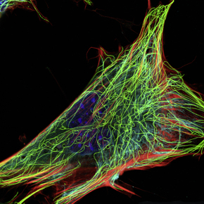
The Leica Microsystems SP8 Laser Scanning Confocal Microscope takes images of structures inside of cells with exceptional clarity. “It is analogous to an MRI that is used in medicine which allows you to see inside the body, but instead of using magnetic resonance the confocal allow us to take ‘optical sections’ through a specimen,” said Dr. Scott Ferguson.
These sections can be reassembled in a computer to generate a 3D representation of the cell and the structures therein.
Though in use for just a couple months, the microscope has generated exciting data both in Ferguson’s research lab as well as the in the Molecular Genetics lab that he teaches. It will also serve as a key instrument in a major research project to be funded by the National Science Foundation. Dr. Scott Medler is also utilizing the microscope in his research into skeletal muscle physiology and the effects of exercise on muscle fiber types.
Ferguson will demonstrate use of the microscope during the reception at 2:15 and 3:15 p.m. in room 115.
The microscope, which carries a price tag of $311,000, was purchased using money from a dedicated equipment budget that is part of the science center’s construction fund.